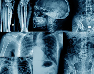
X-rays are a type of high-energy electromagnetic radiation, also called X-rays. X-rays are useful for a variety of purposes including identifying fractures or fractures, ailments, and even allowing security guards to find people’s hidden weapons as they pass through security checkpoints. The scientist who discovered X-rays was Wilhelm Roentgen in 1895. The first X-rays were taken on one of his wife’s hands, including her wedding ring. X-rays are very useful in medicine and are not only the oldest, but also the most widely used type of imaging. Too much energy for X-rays can be harmful to your health. When Wilhelm Röntgen mentioned the X-ray process, he meant that X represented the unknown because the technology fascinated him so much.
The “X” in X-ray stands for the unknown, just as x stands for an unknown quantity in mathematics.
Wilhelm Roentgen discovered X-rays by accident. He was experimenting with some vacuum tubes when he made the discovery.
During World War I, X-rays were already being used for medical purposes, including locating bullets in the human body.
Wilhelm Roentgen was awarded the Nobel Prize in Physics for his invention in 1901.
X-rays were originally considered completely safe to the body, even though x-ray technicians would often suffer burns.
Wilhelm’s wife was not impressed by her husband’s invention. After seeing the image of her hand she said “I have seen my death.”
The first person to die from x-ray radiation exposure was Thomas Edison’s assistant, Clarence Dally, who had worked extensively with X-rays. He died of skin cancer in 1904.
The X-ray has led to many advances in medicine, and its inventor Wilhelm refused to take out a patent because he wanted everyone to benefit from its use.
X-ray technology can be used for elemental analysis and chemical analysis, determining the materials and layers in art objects, buildings, archaeological finds, and more.
X-ray refers to the method used to obtain the image and to the image itself.
Diamonds don’t show up on X-rays.
X-ray is spelled many different ways including xray, X ray, X-ray, and x-ray.
X-rays and radio waves (all electromagnetic radiation) travel at the speed of light in a vacuum (186,000 miles/second).
Wilhelm Roentgen used a zinc box and lead box, when taking x-rays. He did this to protect photographic plates also stored in his lab from being destroyed. He was unaware that this protected him as well. Too much exposure to X-rays can cause cancer.
X-rays have discovered very strange items in pets and human bodies, including pennies and socks swallowed by dogs, gun pellets that were accidentally swallowed, or all manner of awful items puncturing the skull or abdomen.
Scientists that were not yet aware of the dangers of exposure to X-rays often suffered burns, radiation sickness, loss of hair, and cancers.
In a 2009 Science Museum of London poll, the X-ray was voted the most important modern scientific discovery.
Clarence Dally is the first person known to have died from exposure to X-rays. He worked on Thomas Edison’s X-ray light bulb for many years and developed cancerous lesions. This resulted in the amputation of both hands, and his early death at only 39.
X-rays can be divided into hard X-rays and soft X-rays.
As x-rays pass through the body some of the waves are blocked and some pass through, allowing certain images to appear.
In the early 1950s, a British researcher name Rosalind Franklin took the x-ray photos that first showed DNA’s structure, but died before she could share the Nobel Prize with the men more generally given credit for discovering the shape of the “secret of life”—James Watson and Francis Crick.
There are a variety of X-rays used today in addition to the ones used for identifying broken bones and fractures, including contrast x-rays, CT scans, fluoroscopy, dental x-rays, mammograms.
Barium is a substance that is often used with X-rays.
Ultrasound and MRI imaging techniques are not X-ray methods and do not expose the patient to the level of radiation of an X-ray.
X-rays helped us learn more about the structure of DNA and its helix shape.
Because X-rays can be dangerous to a developing fetus, pregnant women are not supposed to have X-rays. Exposure during pregnancy can lead to birth defects and childhood leukemia.
X-rays allowed doctors to see shadows and spots on the lungs of the patient which were caused by the tuberculosis bacteria.
X-rays are known carcinogens. However they can be helpful in medical diagnosis and treatment.
William Roentgen did not patent the X-ray.
It is estimated that approximately 0.4% of the cancers in the United States are due to CT scans.
Some doctors used x-rays to burn off moles and growths on the patient’s skin.
At one time in the 1920s people tried to use X-rays to remove unwanted hair. It was eventually banned by the FDA because it resulted in serious health issues such as cancer.
X-Ray FAQs:
X-rays are a common medical imaging technique used to diagnose and monitor various conditions. Here are some of the most frequently asked questions about X-rays:
1. What is an X-ray?
An X-ray is a type of electromagnetic radiation similar to light, but with shorter wavelengths and higher energy. This allows X-rays to pass through soft tissues like muscles and organs, but they are absorbed by denser tissues like bones. When an X-ray machine directs a beam of X-rays at the body, a special plate or detector captures an image based on how much radiation passes through. Denser tissues appear white on the X-ray image, while softer tissues appear gray or black.
2. What are X-rays used for?
X-rays are a versatile tool used for various diagnostic purposes. Here are some common applications:
- Fractures: X-rays are the primary imaging technique used to diagnose broken bones.
- Joint problems: X-rays can reveal arthritis, joint dislocations, and other abnormalities.
- Chest infections: X-rays can help identify pneumonia, inflammation, or fluid buildup in the lungs.
- Digestive issues: X-rays may be used to examine the digestive tract for blockages, ulcers, or other problems, often with the help of a contrast medium (a special dye).
- Dental problems: X-rays are used to examine teeth and jawbones for cavities, abscesses, or other issues.
3. Are X-rays safe?
X-rays do involve exposure to ionizing radiation, which carries a small risk of damaging cells. However, the amount of radiation used in a typical X-ray is very low and generally considered safe. The benefits of an X-ray in diagnosing a medical condition usually far outweigh the minimal risk.
4. Who should not have X-rays?
While generally safe, X-rays might be avoided in certain situations:
- Pregnancy: Radiation exposure during pregnancy can be harmful to the developing fetus. If an X-ray is deemed necessary, precautions like shielding the abdomen will be taken.
- People who have had multiple X-rays: Repeated exposure to radiation increases the potential risk, so doctors will weigh the necessity of an X-ray against the cumulative radiation exposure.
5. What can I expect during an X-ray?
An X-ray is a relatively simple procedure. Here’s a general idea:
- You will be asked to remove any clothing or jewelry that might interfere with the X-ray image.
- You may need to wear a lead apron to protect certain parts of your body, particularly those not being imaged.
- A technologist will position you based on the area being X-rayed.
- You may be asked to hold still or take a specific breath during the X-ray, which typically takes just a few seconds.
- The entire process is usually completed within 15 minutes.
6. What happens after an X-ray?
The X-ray images will be interpreted by a radiologist, a doctor specializing in medical imaging. The radiologist will send a report to your doctor, who will discuss the results with you and determine the next steps.
7. Are there any alternatives to X-rays?
In some cases, other imaging techniques may be used instead of X-rays, depending on the specific situation. These alternatives might include:
- Ultrasound: Uses sound waves to create images, safe for pregnant women.
- MRI (Magnetic Resonance Imaging): Uses strong magnets and radio waves to create detailed images but may not be suitable for people with certain medical implants.
- CT scan (Computed Tomography): Uses multiple X-ray images to create detailed cross-sectional views, involves slightly higher radiation exposure than a standard X-ray.
Your doctor will recommend the most appropriate imaging technique based on your individual needs.


