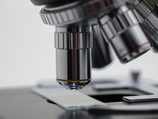
A microscope is a tool that can be used to make very small (microscopic) objects visible to the human eye through magnification. The first microscopes had only one lens and were called simple microscopes. Compound microscopes have at least two lenses and were invented in the 1590’s. The first microscopes relied on light to see the specimen in question and were called optical microscopes. As science advances, new methods are used and today there are a variety of microscopes including the electron microscope, ultramicroscope, and scanning probe microscope.
In 2008 the TEAM 0.5 debuted. It is the world’s most powerful transmission electron microscope and is capable of producing images half a ten-billionth of a meter.
Before the invention of microscopes people believed that illnesses were the result of poisonous gases or evil spirits. Once the microscope was created and people could see viruses and bacteria, these beliefs began to change.
The most common kind of microscope is the compound light microscope.
The very first microscopes were used to study insects, and they were nicknamed ‘flea glasses’.
In a compound light microscope, the object is illuminated: light is thrown on it.
Many believe that Zacharias and Han Jansen created the first compound microscope in the 1590s, but others believe it was Cornelis Drebbel in the 1620s.
The second most common kind are a few kinds of electron microscopes.
In 1625 the name ‘microscope’ was chosen by Giovanni Faber to reference the compound microscope created by Galileo Galilei’s.
Transmission electron microscopes (TEMs) fire cathode rays into the object being looked at. This carries information about how the object looks into a magnetic “lens”.
A compound microscope has at least two lenses, including one at the eye called the eye piece, and one at the end closest to the sample called the objective.
A fluorescence microscope is a special kind of light microscope. In 2014, the Nobel Prize in Chemistry was awarded to Eric Betzig, William Moerner, and Stefan Hell for “the development of super-resolved fluorescence microscopy”.
In 1665 a man named Robert Hooke published a book that included images (hand-drawn) of samples seen under the microscope’s lens. His book was titled Micrographia.
Compound Microscopes are also known as High Power or Biological microscopes.
A microscope works because it is able to distinguish close structures as separate structures.
Compound Microscopes typically, include 3-5 objective lenses that range from 4x-100x. Combined with 10x eyepieces, total magnification ranges from 40x-1000x.
Robert Hooke is credited with the discovery of cells, after studying a cork under the microscope.
Stains are useful for enhancing contrast in the specimen and, therefore, in improving the viewing experience.
Taste buds and red blood cells were identified by Marcello Marpighi. He is known as the father of microscopic anatomy.
There are two broad categories of light microscope: Stereo and Compound. Stereo are Low Power or Dissecting microscopes. Compound are High Power or Biological microscopes.
Antony van Leeuwenhoek invented a single lens microscope in the 1660s that could magnify a sample 200 times.
The magnification power of a light microscope is achieved by multiplying the power of the eyepieces by the power of the respective objective lens.
Antony van Leeuwenhoek is credited with discovering cells in plant tissue, and in animal and human blood and tissue.
Stereo microscopes are used for macro specimens that are visible to the naked eye (insects, crysatsl, pcb etc).
Robert Koch, a German physician and microbiologist, is credited with discovering cholera bacilli and tuberculosis.
A digital microscope can be achieved by simply adding a digital microscope camera to a standard microscope – while retaining maximum flexibility.
Compound microscopes today are so advanced that they can magnify a sample as many as 1000 times.
The resolution of a digital microscope camera is limited by the resolution of your computer monitor.
When a sample under the microscope is photographed (the magnified image) it is called a micrograph.
Most digital microscopes and microscope cameras include basic image capture and documentation software.
An electron microscope uses electrons instead of light to create the magnified image. The first electron microscope was the transmission electronic microscope, invented in 1931 by Ernst Ruska.
Binocular microscopes have a prism, which is either in the microscope head or body tube.
The scanning electron microscope was invented in 1935 by Max Knoll.
The highest resolution microscope measures up to 0.39 ångströms, achieved by researchers at Cornell University (USA), in Cornell University, Ithaca, USA, as published on 18 July 2018. 0.39 ångströms is 3.9X10^-11 metres.
The scanning probe microscope was created in the 1980s, by Gerd Binnig and Heinrich Rohrer.
Lawrence Berkeley National Labs just turned on a $27 million electron microscope. Its ability to make images to a resolution of half the width of a hydrogen atom makes it the most powerful microscope in the world.
The atomic force microscope was created in 1986 by Gerd Bennig.
In 700 BCE the “Nimrud lens,” or rock crystal with a convex shape is believed to have been used as a magnifying lens.
A 500 nanometer long object was the smallest sample seen through a light microscope.
During the year of 167 BCE, there is record of the Chinese culture utilizing the first simple microscope technology. The microscope was made of a lens and a water-filled tube.
When preparing a sample to view under a microscope, sometimes it is stained to make it more visible.
After a lull of excitement, in 1825, Joseph Jackson Lister developed combined lenses.
Microscopes are used for a variety of purposes. They are used for medical purposes, in the diagnosis of illnesses.
The first practical binocular microscope was created sometime during the 1850s at Tulane University.
They are used for biology research, scientific research, medical research, and in environmental scientific research.
In 1957, Marvin Minsky of MIT invented the confocal microscope, a predecessor to today’s confocal laser scanning microscope.