Sonography, a medical imaging technique employing ultrasound waves, has undergone a remarkable evolution since its inception. From its pioneering days in the mid-20th century to the current era of advanced technology and innovation, sonography has significantly impacted medical diagnostics and patient care. This journey encompasses groundbreaking inventions, clinical applications, and technological breakthroughs that have transformed the landscape of medical imaging. In this exploration of 25 historical facts and numerical trivia about sonography, we delve into key milestones, significant figures, and technological advancements that have shaped the narrative of this vital medical field. From its role in obstetrics to its applications in space, sonography continues to push the boundaries of medical science, ushering in new possibilities and contributing to the advancement of healthcare.
Invention and Early Development: Sonography, or ultrasound imaging, traces its roots back to the early 20th century. It was in 1942 that Austrian neurologist Karl Dussik first attempted to use ultrasound to investigate the human brain. The breakthrough came in 1956 when British obstetrician Ian Donald and engineer Tom Brown developed the first practical ultrasound machine. This pivotal moment marked the beginning of diagnostic ultrasound, revolutionizing medical imaging.
First Clinical Ultrasound: In 1958, the first clinical use of ultrasound in obstetrics took place in Scotland, confirming the viability of using this technology for examining pregnancies. The event is noteworthy for pioneering a non-invasive method for observing fetal development and detecting potential complications, fundamentally altering prenatal care.
Significance of 1970s: The 1970s witnessed significant advancements in ultrasound technology. Real-time imaging, introduced in 1972, enabled dynamic visualization of internal structures, facilitating more accurate and detailed diagnoses. This era also saw the emergence of Doppler ultrasound, a technique for assessing blood flow, adding a new dimension to diagnostic capabilities.
First 3D Ultrasound Image: The first 3D ultrasound image was produced in 1987 by Olaf von Ramm and Stephen Smith at Duke University. This groundbreaking development allowed for three-dimensional visualization of the fetus in utero, providing a more comprehensive understanding of anatomy and aiding in the diagnosis of congenital abnormalities.
Ultrasound in Cardiology: By the late 1970s, echocardiography became a crucial application of ultrasound. Dr. Inge Edler and Dr. Hellmuth Hertz conducted pioneering work in cardiac ultrasound, leading to its widespread use in diagnosing and monitoring heart conditions. This marked a turning point in cardiology, providing non-invasive insights into cardiac structure and function.
Portable Ultrasound: In 1985, a significant milestone was achieved with the development of the first portable ultrasound machine by Siemens. This innovation drastically increased accessibility to ultrasound technology, allowing for point-of-care imaging and transforming the dynamics of patient care in various medical settings.
Sonography in Interventional Procedures: The 1990s saw an expansion of ultrasound applications into interventional procedures. Surgeons began using ultrasound guidance for minimally invasive surgeries, biopsies, and drainage procedures, reducing the need for more invasive methods and enhancing precision in medical interventions.
High-Frequency Ultrasound: Advances in transducer technology in the 2000s led to the development of high-frequency ultrasound. Operating at frequencies exceeding 20 MHz, these systems enabled unprecedented resolution, particularly in superficial imaging. This was particularly valuable in dermatology and ophthalmology, where fine structures could be examined with exceptional detail.
Ultrasound in Emergency Medicine: Over the last two decades, ultrasound has become an integral tool in emergency medicine. Rapid, accurate diagnostic capabilities have made it indispensable for assessing trauma, abdominal pain, and other critical conditions in emergency situations, influencing timely and effective medical decisions.
3D Printing from Ultrasound Data: In recent years, the convergence of ultrasound imaging and 3D printing technology has opened new frontiers in medical visualization. Clinicians can now create tangible, patient-specific models from ultrasound data, enhancing pre-surgical planning and education. This integration signifies a remarkable intersection of cutting-edge technologies for improved patient outcomes.
Fetal Gender Determination: Initially, ultrasound was primarily used for medical purposes, but by the 1980s, the technology became a popular tool for determining fetal gender during pregnancy. This non-invasive method, often known as the “gender reveal” ultrasound, gained cultural significance, influencing how families anticipate and celebrate the arrival of a new family member.
Ultrasound Elastography: In the early 2000s, elastography emerged as a novel technique within ultrasound imaging. This method assesses tissue stiffness, providing valuable information for the diagnosis of liver fibrosis and other conditions. The introduction of elastography marked a significant step in non-invasive assessment, reducing the need for more invasive procedures like liver biopsies.
Contrast-Enhanced Ultrasound (CEUS): The integration of contrast agents in ultrasound, known as Contrast-Enhanced Ultrasound (CEUS), became a notable development in the 1990s. By improving visualization of blood flow and organ perfusion, CEUS enhanced diagnostic capabilities in fields such as oncology and hepatology, where detailed vascular information is crucial for accurate diagnosis and treatment planning.
Focused Assessment with Sonography for Trauma (FAST): In the 1990s, the FAST exam gained prominence in emergency medicine. This focused ultrasound assessment aids in the rapid evaluation of trauma patients, specifically detecting free fluid in the abdominal and chest cavities. The FAST exam significantly contributed to timely decision-making in trauma care and has become a standard tool in emergency departments worldwide.
Telemedicine and Telesonography: With the advent of digital technology, the 21st century witnessed the integration of telemedicine into ultrasound practices. Telesonography allows remote interpretation of ultrasound images and consultations, overcoming geographical barriers and improving access to specialized medical expertise, especially in underserved areas.
AI and Automation in Sonography: In recent years, artificial intelligence (AI) has entered the realm of sonography, offering automated image analysis and interpretation. AI algorithms aid in the detection of abnormalities, improving diagnostic accuracy and efficiency. This development represents a paradigm shift in how ultrasound data is processed and interpreted, potentially revolutionizing diagnostic workflows.
Endoscopic Ultrasound (EUS): Combining endoscopy and ultrasound, Endoscopic Ultrasound (EUS) became a significant advancement in the 1980s. This technique allows for detailed imaging of the gastrointestinal tract and adjacent structures, providing valuable information for the diagnosis and staging of various digestive system disorders.
Ultrasound-Guided Nerve Blocks: The application of ultrasound in regional anesthesia gained momentum in the 21st century. Ultrasound-guided nerve blocks enable precise targeting of nerves for anesthesia, reducing complications and improving the safety and efficacy of regional anesthesia procedures.
Ultrasound in Sports Medicine: Ultrasound has found extensive use in sports medicine for the assessment of musculoskeletal injuries. This includes the evaluation of soft tissues, tendons, and ligaments, allowing for accurate diagnosis and targeted treatment in athletes. The real-time imaging capabilities of ultrasound contribute to effective rehabilitation strategies.
Portable 4D Ultrasound: Advancements in technology in the 2010s led to the development of portable 4D ultrasound machines. These compact, high-performance devices offer real-time 3D imaging, enhancing the visualization of dynamic processes in the body. Portable 4D ultrasound has proven valuable in diverse medical settings, including obstetrics, emergency care, and point-of-care applications.
Ultrasound in Neurology: Over the last decade, ultrasound has gained traction in neurology for assessing various conditions, including neurodegenerative diseases and cerebrovascular disorders. Transcranial Doppler ultrasound, in particular, allows non-invasive monitoring of blood flow in the brain, aiding in the diagnosis and management of neurological disorders.
Sonography in Veterinary Medicine: Beyond human medicine, ultrasound has become an indispensable tool in veterinary practice. Veterinary sonography allows for the imaging of animals’ internal structures, aiding in the diagnosis of conditions ranging from pregnancy to abdominal abnormalities in pets and livestock.
Portable Ultrasound in Disaster Response: Portable ultrasound devices have played a crucial role in disaster response scenarios. Their mobility and ease of use make them valuable tools for rapid medical assessments in the aftermath of natural disasters or emergencies, facilitating timely and efficient medical interventions.
Ultrasound in Space: Ultrasound technology has even found applications beyond Earth. In space exploration, portable ultrasound devices have been utilized aboard spacecraft to monitor astronauts’ health, providing a means for non-invasive medical evaluations during long-duration space missions. This illustrates the adaptability and versatility of ultrasound technology in diverse environments.
Holographic Ultrasound Displays: The cutting-edge developments in ultrasound technology include the exploration of holographic ultrasound displays. This futuristic approach aims to transform the way medical professionals visualize and interact with ultrasound data, potentially enhancing diagnostic precision and educational experiences in the field of medical imaging. Although still in the experimental stage, holographic ultrasound displays represent an exciting frontier in the evolution of ultrasound technology.
People often ask:
What is sonography done for?
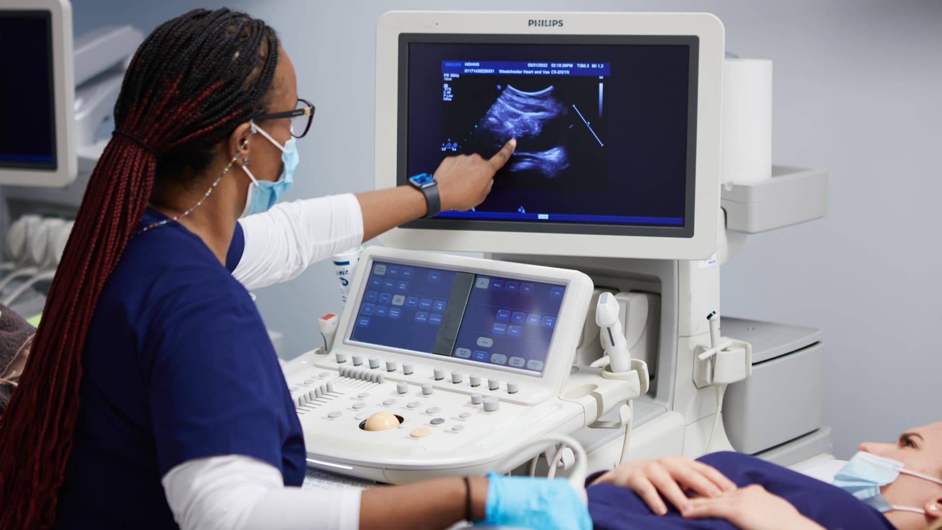
Sonography, also known as ultrasound imaging, is primarily employed for non-invasive visualization and assessment of internal body structures. It utilizes high-frequency sound waves to produce real-time images of organs, tissues, and blood flow. Common applications include diagnosing and monitoring conditions in various medical specialties such as obstetrics, cardiology, gastroenterology, and musculoskeletal imaging. Sonography is instrumental in identifying abnormalities, guiding medical procedures, and providing valuable insights for medical professionals in different fields.
What is the difference between ultrasound and sonography?
The terms “ultrasound” and “sonography” are often used interchangeably, but there is a subtle distinction. “Ultrasound” refers to the technology itself, involving the use of high-frequency sound waves for imaging. On the other hand, “sonography” refers to the actual procedure or technique of creating images using ultrasound. In essence, ultrasound is the technology, while sonography is the application of that technology to produce diagnostic images.
What does the sonographer do?
A sonographer is a skilled healthcare professional trained to perform sonography. The sonographer operates the ultrasound equipment, capturing images and sometimes recording measurements for interpretation by a radiologist, obstetrician, or other specialized healthcare providers. They play a crucial role in patient care, ensuring the quality of images, providing support and reassurance to patients during the procedure, and collaborating with the medical team for accurate diagnosis and treatment planning.
What is sonography in pregnancy?
In pregnancy, sonography is extensively used for monitoring fetal development and assessing the health of both the mother and the unborn child. Obstetric sonography involves the use of ultrasound to visualize the fetus in utero. It helps determine gestational age, detect multiple pregnancies, assess fetal growth and anatomy, and identify potential complications such as placental abnormalities or birth defects. Additionally, sonography is employed for procedures like the nuchal translucency test and Doppler ultrasound to evaluate blood flow in the umbilical cord and other fetal vessels. Overall, sonography in pregnancy is a crucial tool for ensuring the well-being of both the expectant mother and the developing fetus.